Artist Challenge – Day 3

3D, Anatomy, Graphical Works, Molecular

For day 3 I wanted to go back and look at some of my scientific 3D work.  Sometimes the stuff you didn’t think so much about in the moment, looks better when you go back to it.

<a href="https://kulaga.wordpress.com/wp-content/uploads/2012/08/ambientulcer.jpg”>AmbientUlcer

HIV Reverse Transcriptase with RNA/DNA

HIV Reverse Transcriptase with RNA/DNA
pdb model 2hmi

Platelet1


I was nominated by the talented Briana Hertzog to post 3 images a day for 5 days.

Today I nominate my VCU  and UIC partner in crime Jennifer Rogers !! I also nominate another BVIS alumi who has magical work and was amazing support for our two years in grad school, Natalie Doolittle. ♡

3D Heart

3D, Anatomy, Crafty


About 2 years ago I needed to make a 3D model of the human heart for my research project.
3D anatomical heart

Then about a month ago I decided to try and 3D print it. The models are SUPER small ( 0.454w x 0.83d x 0.507(in) ). The following two objects are test prints of the model in strong flexible white plastic (left) and metallic plastic (right).


3D printed heartsThe models were printed via Shapeways, and the plan was to make necklaces for Valentines Day, but I think I will make miniature wall plaques instead. I will redo the model to create a point to hang the hearts from for the necklaces, and make sure the walls are thicker. I don’t want to mess these precious little hearts up!

Feel free to visit the model’s site here: http://shpws.me/A9oF .  While I don’t think others can print the model, because some of the walls are thin, everything seemed to turn out great! Some of the details (vessels)  printed a little messy since they are thinner than spec. But I like it, it gives the heart a more organic feel. ❤

Microbial Magic

Anatomy, Inspiration, Prints, Traditional Work

The season of giving will soon be upon us so I thought I this would be a great opportunity to search the web and curate a mini exhibition of the anatomically awesome things that people create! For the first of this series lets start small, with some microbes!

Society6 is one of the sites I joined last year to help promote my work. And through it I have been able to find a variety of fresh yet talented artists:

Microbe 1 by Veronica Martinis


Microbe Family by AMOSLIDE

(And I am not quite sure what this is, but I like it too  … )

Microbes are also quite fashionable:

Bacteria – Thousands Of ‘Em (Pink) by Chayground

Microbe Onsie

Histoplasma Capsulatum by Yours Truely 😉 ( Autumn Kulaga )

Retro Amoeba Earrings by LemantulaDesigns


Or maybe you know someone that appreciates more tangible objects. You know grown adults searching for “collectables” ( a.k.a desk toys) .

Bacteriophage T7 Model

Shapeways is an amazing site bringing 3D printing and prototyping capability to the masses.

4c9o by Virtox

E-coli Toxin by Anthromod


And of course we can’t forget about the more commonly known plush microbes. Cuddly enough for an infant, even the bed bug is adorable!

Bed Bug Plush


Alicia Watkins is an Etsy Vendor who will create any one of a variety of microbes by cross stitch:

Microbes cross stitch set by Alicia Watkins

Or perhaps you are crafty enough to try out some of her patterns yourself?!


Know of any other microbial wonders? Feel free to share in the comments of this post!

Classical Anatomy

Anatomy, Inspiration

In these stunning sculptures Cao Hui mixes anatomical realism with classical art. Think Michelangelo meets Eycleshymer & Schoemaker 😉

“Using Inspired by Alexandros of Antioch‘s Venus de Milo, Cao Hui‘s reinterpretation of the classic piece cleverly infuses the artist’s own affinity for Photorealism, Realism, and Hyperrealism. Hui is well known for his fleshy masterpieces and often uses his work to play tricks on the naked eye.” – QuietLunch

CaoHui1 CaoHui5Cao-Hui-2

Juxtapoz.com’s Feature on Cao Hui


cao-hui_02
cao-hui-sculpture-04 caoChair CaoHui31


Anatomy with CSS 3D

Anatomy, Inspiration

This evening I want to share with you this post I came across while doing research of webGL frameworks. What I also found is this slider which allows for the display of pre-rendered images from volume rendering. It wowed me…

CSS 3D CSS 3D MRI

” I geeked out by applying fake specular lighting, for that ‘fresh meat’ look, and volumetric obscurance to enhance the sense of depth on the inside.” – @unconed
http://acko.net/blog/this-is-your-brain-on-css/

Documentation

3D, Anatomy

Documenting your work is just as important as creating work. Lately I find myself working more to refine pieces and figuring out how to display it, in order to share what I have created. So to show for this I have two new videos!

The above is my Blood Components Interactive. With the help of JD Pirtle and others in the UIC – EVL lab, I now have some amazing footage of this application! This interactive was built on OmegaDesk with the help of Victor Mateevitsi,  Alessandro Febretti & Arthur Nishimoto.  The environment I developed for included a 3D viewing screen, a 2D touch screen and Kinect for gestural interaction.  My personal objective was to explore designing an application which involved different types of interaction  I wanted to think about how you would display 3D assets in this environment and also how to share textual information in the same context. I choose blood components because I like working with cellular and molecular structures. I see them as abstract characters, and while it is a serious subject, you can have a lot of fun with them!  I also enjoyed creating the flow of the blood stream. While the irregular movement of the objects can make it difficult at first, I imagine kids would enjoy the challenge of going after the different floating cells.  Ideally I see this interactive going in a science or health museum which showcases immunity.

Next I would like to share the video I made using mMaya’s beta-factor to animate the movement of HIV-RT CPK molecular representation. The main star of the animation is HIV’s Reverse Transcriptase (HIV-RT). This little devil travels with HIV’s viral RNA, turning it into DNA which is then integrated into the host cell by HIV Integrase.

This semester I am finishing up research, creating a game in Unity with teammates, redoing my website, and creating an animation in Maya. I hope you will enjoy what is to come!

For a short time you can also visit game.encephalostudios.com for a quick peak at a montage of my work.

An eventful spring is ahead!

Anatomy, Crafty, Events

To start off the semester I want to tell you about the great things we are doing this Spring!  We have three great events coming up soon.  Today I will inform you about ‘Anatomical Love’, ‘Digital Creations’ & our ‘Anatomical Zine’!

Next week the students of BVIS are hosting a gallery exhibition at Pilsen Thrift.

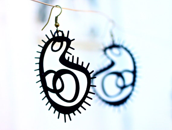
Anatomical Love will be an amazing show highlighting some work created by students. On display will be work completed while in the program as well as personal artwork. There are even whispers that some of the artwork may be for sale?!

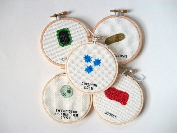
Here is a sneak peak of my cross-stitching project:

Heart - CrossStitch

(While this masterpiece is quite tedious, it did give me an excuse to watch over 40 hours of The Tudors)

If you can’t make it out February 8th, then come see us March 1st at the National Museum of Health and Medicine in Chicago. The NMH+MC is a private venue, therefore an RSVP is required. Email uicbvis.students@gmail.com with subject “Digital Creations RSVP“, please include your name and name of any guests.

DigitalCreation

Digital Creations is a chance for me and my colleagues to show artwork made with different media while in UIC’s Biomedical Visualization Program. Ranging from 3D medical tools to personalized independent projects this exhibition will highlight the range of work we are capable of.

And last but not least, the Student Association of Medical Artists (SAMA – the BVIS student organization) will be represented at the Chicago Zine Fest.  On March 9th from 11am – 6pm we will have two tables set up, one for a bake sale ( baked good for donations) and another to sell Anatomical Zines.

chicagoZineFest

 This festival is serving as our annual fundraiser. Profits will be used to help fund figure drawing held throughout the year, as well as reimburse students who submit there work to the Salon at the Association of Medical Illustrators Annual Meeting ( a very prestigious event for our field 😉 ).

We can’t wait to see you this semester!

Cervical Spinal Fusion of C1 & C2 Vertebrae

Anatomy, Surgical, Traditional Work

The surgery I observed was to correct a type II fracture of the dens on the axis, a bone articulating with the atlas.  The atlas and the axis are the first vertebrae of the spine. The atlas (C1) articulates with the base of the skull. This atlantooccipital joint helps create a head nod (‘yes’ motion). The atlantoaxial articulation rotates along a vertical axis, creating a head shake (‘no’ motion).

The patient had broken their dens, a process on the axis articulating with the atlas. Even though the neck had been braced for some time, the area would not heal. To correct this screws and rods were placed in the pedicle of the vertebra. Being that they are irregular vertebrae, it might be more correct to say the screws were under the articulating facets. This is lateral to the vertebral foramen and away from the vertebral arteries.


For my portfolio I plan to finish filling in the tone of the remaining drawings but for the assignment the first three illustrations were fully rendered in tone. I really like using the toned background.  I think it was a good choice, especially since I had so much bone to render! The toned paper gave me more flexibility when rendering the light tones.


I can’t wait to fill in the bone chips and render the suture line!

Sketching in the OR

Anatomy, Surgical, Traditional Work

While some people might find being in a surgical operating room uneasy and slightly awkward, I find it amazingly intriguing. First of all you get to wear and sweet get up:

Stylish… I know, but it’s all in the name of hygiene  For our first surgical illustration assignment the goal was to view as many surgeries as possible and see different techniques. This exercise was to help build our surgical illustration visual library, a mental reference of as many tissue manipulations as possible.

Deep incision on the upper arm

Blunt dissection of superficial tissue

When you first go in the OR, anything can be happening. Sometimes no one is in the room, at others the surgery has already started. Everyone has a face mask on, so you can only see eyes. I found it took a little bit of re-learning body language without certain facial features to pick up on. But you quickly pick up on interpretations as well as etiquette.

Tissue manipulation and suture hemostasis; removal of fistula

Cauterization of skin tissue using hemostat

Some surgeries I couldn’t see anything, I would be on my tippy-toes, or crouching down just to get a glimpse of what was going on. If you have a more inviting OR team, they will help direct you to a place where you can see something, but still be out of the way. At other times the surgical field is so small, that regardless of how people move, you do not see anything. In instances like this I find it helpful to stay close ( but out of the sterile field) and listen to any discussion about the procedure.

Suturing skin to areola and staple retraction

Suturing scalp

Cauterizing skin tissue (hemostasis)

I am very thankful for this opportunity. It is amazing to be able to observe some of the complicated procedures surgeons are performing in real time. The chance to see teams of people work together for the health of one individual.